In diagnostic medicine, biopsies, where a sample of tissue is extracted for analysis, is a common tool for the detection of many conditions. But this approach has several drawbacks – it can be painful, doesn’t always extract the diseased tissue, and can only be used in a sufficiently advanced disease stage, making it, in some cases, too late for intervention. These concerns have encouraged researchers to find less invasive and more accurate options for diagnoses.
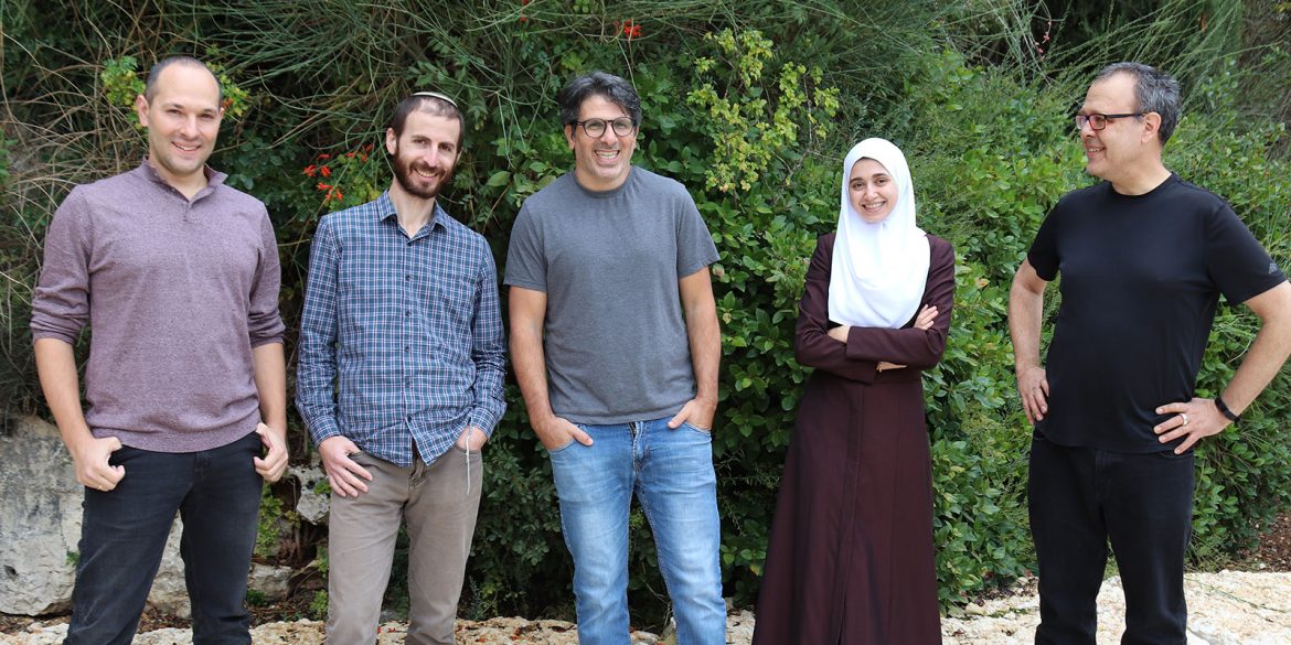
Professor Nir Friedman and Dr. Ronen Sadeh of the Life Sciences Institute and School of Computer Engineering have published a study in Nature Biotechnology that shows how a wide range of diseases can be detected through a simple blood test. The test allows lab technicians to identify and determine the state of the dead cells throughout the body and thus diagnose various diseases including cancers and diseases of the heart and liver. The test is even able to identify specific markers that may differ between patients suffering from the same types of tumorous growths, a feature that has the potential to help physicians develop personalized treatments for individual patients.
The test relies on a natural process whereby every day millions of cells in our body die and are replaced by new cells. When cells die, their DNA is fragmented and some of these DNA fragments reach the blood and can be detected by DNA sequencing methods. However, all our cells have the same DNA sequence, and thus simply sequencing the DNA cannot identify from which cells it originated. While the DNA sequence is identical between cells, the way the DNA is organized in the cell is substantially different. The DNA is packaged into nucleosomes, small repeating structures that contain specialized proteins called histones. On the histone proteins, the cells write a unique chemical code that can tell us the identity of the cell and even the biological and pathological processes that are going on within it. In recent years, numerous studies have successfully developed a process where this information can be identified and thus reveal abnormal cell activity.
A new approach advanced by Hebrew University researchers, Professor Friedman and Dr. Ronen Sadeh is able to precisely read this information from DNA in the blood and use it to determine the nature of the disease or tumor, exactly where in the body it’s found and even how far developed it is.
The approach relies on analysis of epigenetic information within the cell, a method which has been increasingly fine-tuned in recent years. “As a result of these scientific advancements, we understood that if this information is maintained within the DNA structure in the blood, we could use that data to determine the tissue source of dead cells and the genes that were active in those very cells. Based on those findings, we can uncover key details about the patient’s health,” Professor Friedman explains. “We are able to better understand why the cells died, whether it’s an infection or cancer and based on that be better positioned to determine how the disease is developing.”
Along with the clear diagnostic benefits of this process, the test is also non-invasive and far less expensive than traditional biopsies. Dr. Ronen Sadeh said, “We hope that this approach will allow for earlier diagnosis of disease and help physicians to treat patients more effectively. Recognizing the potential of this approach and how this technology can be so beneficial for diagnostic and therapeutic purposes, we set up the company Senseera which will be involved with clinical testing in partnership with major pharmaceutical companies with the goal of making this innovative approach available to patients.”
Sadeh, R., Sharkia, I., Fialkoff, G. et al. ChIP-seq of plasma cell-free nucleosomes identifies gene expression programs of the cells of origin.
DOI: 10.1038/s41587-020-00775-6.
Full article available to view here
This work was supported by: European Research Council’s AdG Grants 340712 “ChromatinSys” (NF) and 786575 “RxmiRcanceR” (EG); Israel Science Foundation’s I-CORE program grant 1796/12 (TK and NF) and grants 2612/18 (NF), 3020/20 (AG), 2473/17 (EG), and 486/17 (EG); Israel Ministry of Science and Technology grant 3-14352 (AG); NIH Grants RM1HG006193 (NF) and CA197081-02 (EG); Deutsche Forschungsgemeinschaft (DFG) SFB841 (EG); DKFZ-MOST grant (EG).
Photo credit: The Hebrew University
